

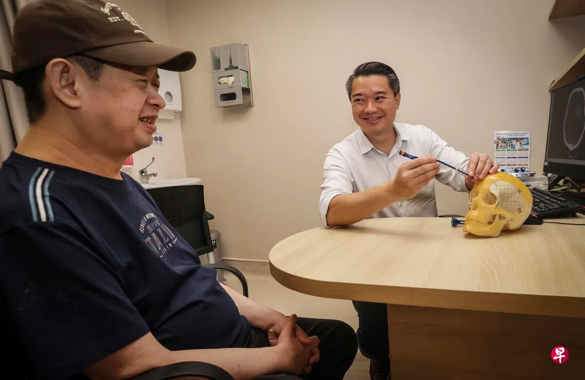
In January this year, 44-year-old Kenneth Yeo suffered a sudden haemorrhagic stroke. By the time he arrived at hospital, he was semi-conscious and paralysed on his right side. Doctors were not optimistic about his prognosis: even if surgery saved his life, he would very likely be bedridden or dependent on a wheelchair for the rest of his life.
But just three days after undergoing minimally invasive brain surgery through a small incision to remove the blood clot, Yeo regained sensation and movement in his previously paralysed right limbs. A month later, he was able to walk steadily out of hospital. During Chinese New Year, he even took public transport with his family to visit relatives and friends.
His sister, Yeo Yilun, a 52-year-old accountant, said he has now made an almost complete recovery and is much as he was before the stroke.
Conventional surgery to remove a blood clot in the brain requires a craniotomy. Doctors make an incision in the scalp and saw an opening about 10 to 15 centimetres wide in the skull. They then open the dura mater and retract brain tissue, sometimes even having to cut through some brain tissue and neural networks to remove the clot and stop the bleeding.
In an interview, Dr Lee Chee Hoe Lester, senior consultant in the Department of Neurosurgery at the National Neuroscience Institute, acknowledged that while conventional surgery can save lives, it carries greater risks because it requires extensive retraction of brain tissue. Depending on the part of the brain affected, patients may experience complications such as brain swelling, seizures, and impaired memory, movement and vision after surgery.
To reduce surgical risks and improve the chances of recovery, the National Neuroscience Institute's neurosurgical team has in recent years been progressively introducing a small-incision technique known as Minimally Invasive Transsulcal Parafascicular Surgery (MIPS) at Changi General Hospital, Singapore General Hospital and Tan Tock Seng Hospital.
Dr Lee said the institute's neurosurgeons have used the minimally invasive technique to treat more than 160 deep-seated brain lesions since 2018. About half of these cases involved brain haemorrhages and surgery to remove blood clots.
Tubular instrument reaches deep into the brain while avoiding vital neural pathways
During the operation, doctors first make a small incision in the patient's scalp and remove a small section of skull measuring only about 3 centimetres in diameter. Using an intraoperative navigation system, they plan the best route based on the patient's CT scans. A tubular instrument is then guided along the brain's natural grooves to reach the deep-seated clot while avoiding important neural pathways.
Once inside the brain, the tube acts as a miniature retractor, gently holding brain tissue apart. The surgeon then uses a microscope to suction out the clot and stop the bleeding. After the operation, the small piece of skull is replaced and secured.
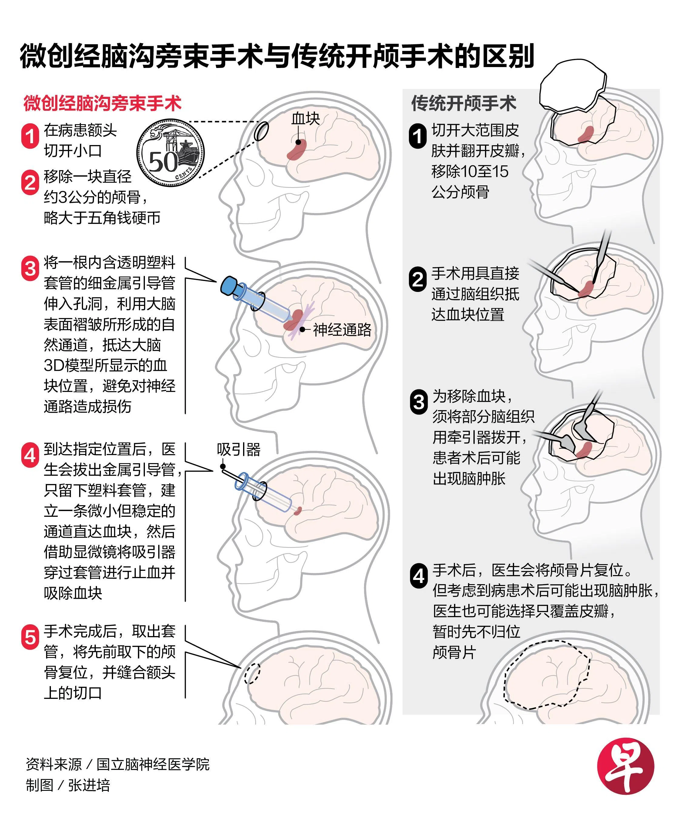
Dr Lee explained that neural pathways are like the brain's “information highways”, carrying signals between different regions of the brain, as well as between the brain and spinal cord. Damage to them can affect a patient's thinking, memory, behaviour, speech and ability to walk. The specific effects depend on which “pathway” is affected.
Compared with conventional surgery, MIPS minimises damage to brain tissue and neural networks, preserving as much of the patient's brain function as possible.
According to a study published by the team this year, MIPS operations performed between 2018 and 2024 took about two hours on average — nearly half the time needed for a conventional craniotomy. This means patients spend less time under general anaesthesia and lose significantly less blood, while a greater proportion of the clot is also removed.
No deaths within six months of surgery; most patients regain ability to care for themselves
In terms of postoperative recovery and survival, patients who underwent MIPS spent about 1.5 fewer days in intensive care on average. There were no deaths in this group within six months of surgery, compared with a mortality rate of about 15% in the conventional craniotomy group.
Patients who underwent minimally invasive surgery were also significantly less likely to have severe disabilities after six months. The vast majority regained the ability to care for themselves, without being bedridden or dependent on a wheelchair in the long term.
Dr Lee said only a few major hospitals in Singapore currently have the necessary equipment and specialist teams, so about 90% of haemorrhagic stroke patients still undergo conventional craniotomies. To benefit more patients, the National Neuroscience Institute is actively promoting the technique among neurosurgeons in Singapore and other countries in the region through training courses.
